
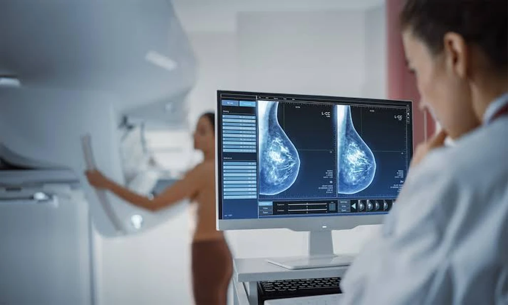
Finding a lump in your breast can trigger immediate anxiety, but the discovery itself doesn’t provide answers. Mammograms and ultrasounds can identify suspicious areas, yet these imaging tools have limitations. They show shapes, densities, and blood flow patterns, but they cannot definitively determine whether tissue is benign or malignant. When imaging results fall into gray areas-marked as BI-RADS 4 or 5 classifications-doctors need cellular evidence to guide treatment decisions.
Why Imaging Alone Cannot Confirm Diagnosis
Breast imaging excels at detection but struggles with certainty. A mammogram might reveal a spiculated mass, which raises concern, but similar patterns can appear in both cancerous tumors and benign conditions like radial scars. Ultrasound can distinguish solid masses from fluid-filled cysts, yet solid lesions require further investigation. MRI offers the highest sensitivity, detecting up to 90% of breast cancers, but it also produces false positives in approximately 10-20% of cases, leading to unnecessary anxiety and procedures.
The problem lies in overlapping characteristics. Fibroadenomas, a common benign tumor in younger women, can appear irregular on imaging. Fat necrosis from previous trauma or surgery can mimic malignancy. Papillomas in milk ducts may show calcifications similar to ductal carcinoma in situ. Without examining actual cells under a microscope, radiologists cannot make definitive diagnoses, and treatment cannot proceed with confidence.
The Role of Tissue Sampling in Breast Health
When imaging identifies a suspicious abnormality, the next step involves obtaining a tissue sample for pathological analysis. This process moves beyond pictures to examine the cellular architecture, identifying specific markers that reveal whether cells are cancerous, precancerous, or benign. The sample allows pathologists to determine tumor grade, hormone receptor status, and HER2 expression-information that directly shapes treatment plans.
Three main approaches exist for tissue sampling. Fine needle aspiration uses a thin needle to extract cells, but it provides limited material and cannot always distinguish between invasive and non-invasive cancer. Surgical excisional biopsy removes the entire lump, which guarantees adequate tissue but requires operating room time, general anesthesia, and leaves visible scarring. The middle approach uses a core needle technique guided by ultrasound or stereotactic mammography, extracting multiple tissue cores through a single small incision. This method provides sufficient material for comprehensive pathology while avoiding surgery for most patients.
How Advanced Facilities Approach Diagnostic Precision
Multi-specialty hospitals in Abu Dhabi have adopted image-guided core needle techniques as the standard for investigating suspicious breast lesions. Cleveland Clinic Abu Dhabi, a multi-specialty hospital providing comprehensive cancer care and diagnostic imaging services across the emirate, offers the breast biopsy procedure using real-time ultrasound guidance. This approach allows radiologists to visualize the needle entering the target tissue, ensuring accurate sampling of the abnormal area while avoiding blood vessels and ensuring patient safety throughout the procedure.
The procedure typically takes 30 to 60 minutes and uses local anesthesia. Patients remain awake but feel only pressure rather than pain. The radiologist obtains multiple core samples-usually five to twelve-to account for tumor heterogeneity, where different parts of the same mass may show varying characteristics. A small marker clip is often placed at the biopsy site, creating a reference point visible on future imaging, which proves valuable if the area requires surgical removal later.
What Patients Should Expect During Recovery
Recovery from core needle biopsy differs substantially from surgical procedures. Most patients return to normal activities within 24 hours, though heavy lifting and vigorous exercise should wait 48 to 72 hours. Bruising occurs in approximately 60% of cases and typically resolves within two weeks. Ice packs during the first 24 hours minimize swelling, while firm support from a well-fitted bra reduces discomfort.
Complications remain rare but possible. Bleeding affects fewer than 1% of patients and usually stops with direct pressure. Infection occurs in less than 0.5% of procedures. Some women experience a vasovagal response during the procedure-feeling lightheaded or nauseated-which resolves quickly when lying flat. The small incision typically measures three to five millimeters and heals with minimal scarring, often becoming invisible within months.
Interpreting Results and Planning Next Steps
Pathology results typically arrive within three to five business days. Benign findings-fibroadenoma, fibrocystic changes, or normal tissue-usually require only continued monitoring with periodic imaging. High-risk lesions such as atypical ductal hyperplasia or lobular carcinoma in situ often prompt surgical excision, as these conditions carry elevated cancer risk and small samples may miss adjacent malignancy.
Malignant results trigger multidisciplinary review involving surgeons, medical oncologists, and radiation oncologists who examine the pathology alongside imaging to stage the cancer and recommend treatment. The biopsy report provides critical information about hormone receptors and growth factor status, determining whether the tumor might respond to hormonal therapy or targeted treatments. This personalized approach has transformed breast cancer care, moving away from one-size-fits-all protocols toward tailored strategies based on tumor biology.
Choosing Where to Have Diagnostic Procedures
The facility performing your biopsy matters significantly. Volume correlates with accuracy-radiologists who perform these procedures regularly develop sharper skills in needle placement and lesion targeting. Equipment quality affects precision, with newer ultrasound machines providing clearer visualization. Turnaround time for pathology results varies by institution, and choosing the right clinic involves evaluating these practical factors alongside credential verification and patient experience.
Integration between diagnostic and treatment services streamlines care when results indicate cancer. Hospitals offering comprehensive cancer programs can move patients directly from diagnosis to treatment planning without referrals to outside facilities. This coordination reduces delays and ensures that the surgical team has direct access to the radiologist’s notes and images, improving surgical planning and outcomes.
The Future of Breast Diagnostics
Emerging technologies promise to refine diagnostic accuracy further. Contrast-enhanced mammography combines traditional imaging with iodinated contrast agents, highlighting areas with increased blood flow characteristic of tumors. Molecular breast imaging uses radioactive tracers that concentrate in cancer cells, detecting lesions that traditional mammography might miss in dense breast tissue. These advances may reduce the number of biopsies needed by improving the specificity of initial imaging.
Some research explores liquid biopsies, which analyze circulating tumor DNA in blood samples. While promising for monitoring cancer recurrence, this technology cannot yet replace tissue sampling for initial diagnosis. The cellular architecture visible under a microscope still provides information that blood tests cannot match. As healthcare systems explore innovative care delivery models, diagnostic procedures like breast biopsy remain anchored in specialized clinical settings where imaging equipment, pathology labs, and expert staff converge.
Moving Forward With Confidence
A suspicious finding on breast imaging creates uncertainty, but modern diagnostic techniques provide clear answers efficiently and safely. Core needle biopsy bridges the gap between imaging concern and treatment clarity, delivering the cellular evidence needed for confident decision-making. While the procedure itself takes less than an hour, the information it provides shapes months or years of medical management. Understanding this process-why it’s necessary, how it works, and what results mean-empowers patients to participate actively in their care, asking informed questions and making decisions aligned with their values and circumstances.
Whether results reveal benign findings requiring only surveillance or indicate cancer necessitating treatment, tissue diagnosis removes ambiguity. In breast health management, precision matters, and biopsy remains the gold standard for achieving it.
